|
پروفسور محمد
حسین سلطان زاده
استاد دانشگاه علوم پزشکی شهید بهشتی
متخصص کودکان ونوزادان
طی دوره بالینی عفونی از میوکلینیک آمریکا
دبیر برگزاری کنفرانس های ماهیانه گروه اطفال
دانشگاه علوم پزشکی شهید بهشتی
|
SHIRVANI F.
MD MS
KALANTAR MOTAMEDI M. MD
SHEIKHOLESLAM H. MD
RADFAR M. MD
به اتفاق اعضای هیئت علمی گروه کودکان
بیمارستان امام حسین
|
CHIEF COMPLAINT: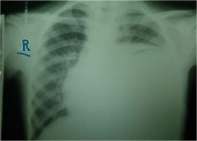
An 8 years
old girl admitted at hospital in 82/01/19 with a chief complaint of fever,
respiratory distress and night sweat
HOPI:
she had a
history of two months of weight loss ,fever and night sweat which was under
antibiotic prescription without significant improvement
She was the
fourth child in the family from a mother G5P5Ab0 and the result of a NVD
without complication.
She was
from afganistan and had a long contact with a patient with TB , she lived near a
sheep raising, no history of hospital admission , or specific illness,
vaccination history was positive.
PH. E.
T= 38 C
ORAL PR=110/Min
RR=33/MIN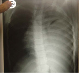
BW=19KG
General
appearance was good .
Head and
neck=normal. No adenopathy
chest =
heart sounds were normal, full dullness on left lower chest and decrease of the
breath sounds was apparent .
Lab
investigations:
CBC DIFF ,
ESR, CRP , BS , CA,ELECTROLYTES ,Urea,Creatinine U/A , U/C , B/C , ABG , ELIZA
IgG AND IgM for echinococcus granulosus
Needle
aspiration of fluid
CHEST X RAY
, CHEST CTSCAN
Abdominal
sonography
WBC
=9100/mm3 poly=54%,lym=44%,Eos=1%,
mono=1%,plt=607000,ESR=123
BS,UREA,CREATININE=NL
NA,K,CA,P,=NL
CRP=3+
U/A , U/C ,
B/C =Neg , ABG=NL
Elisa for
echinococcus IgG AND IgM=Neg
Aspiration
of fluid results: LDH=40,PRO=10,SUGAR=30mg/dl and smear and
culture was negative
CHEST X RAY:
Homogenous
opacity on left lower lobe that obscures the diaphragm and causes the shift of
trachea and heart to the right ,a crecentric shadow is seen at the top of the
opacity that suggests its CYSTIC NATURE, CTscan of thorax was recommended .
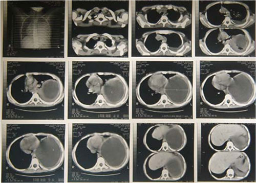
CHEST CTSCAN: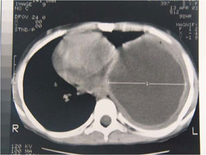
In the
posterior left hemi thorax there was a big cystic mass with thick layer and
pleural thickness , it seems it is an encapsulated empyema.
Abdominal
sonography was normal.
Other
investigations:
PPD=NEG
three times
gastric lavage for BK was negative
What is the patient's possible diagnosis?